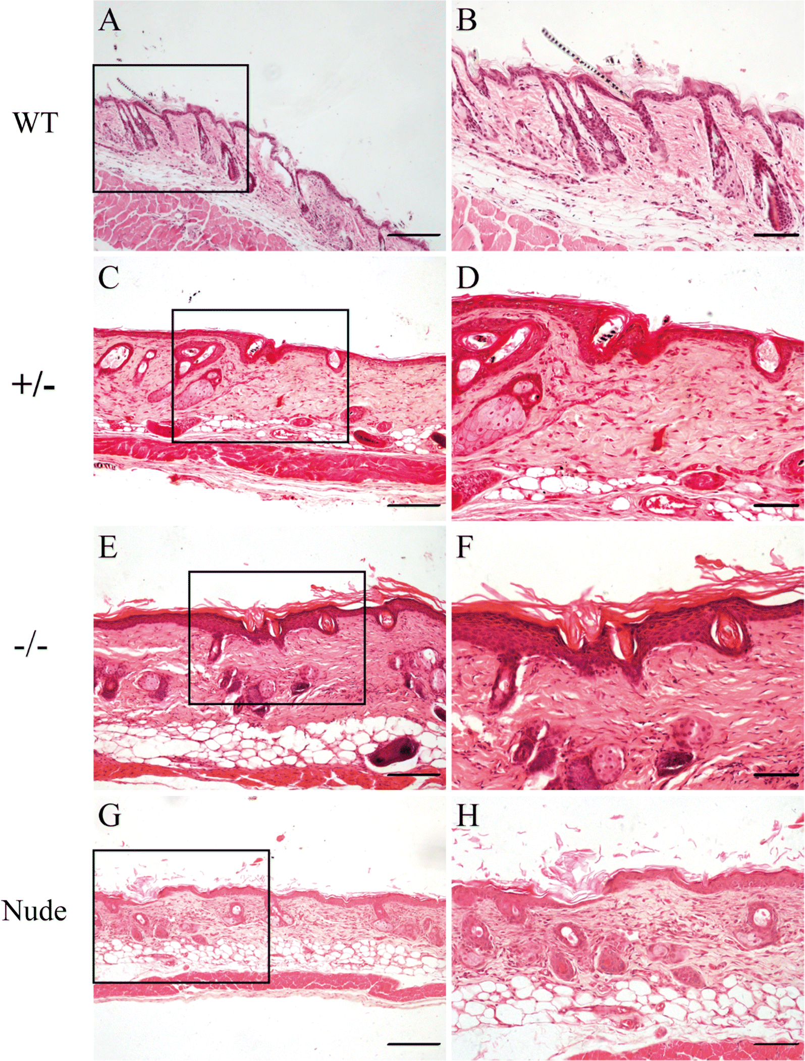
Fig. 5

Histological abnormalities in the skin of PLCD1-deficient mice (F1). H&E staining of dorsal skin sections from wild-type (A, B), heterozygous (±) (C, D) and homozygous (−/−) (E–F) PLCD1-deficient mice, and nude mice (G, H). In B, D, F and H are higher magnifications of the black boxes indicated in A, C, E and G, respectively. Scale bar: Fig. 4A, C, E, G: 100 μm; Fig. 4B, D, F, H: 50 μm